


CYANOSIS IN BROILER BREEDER MALES
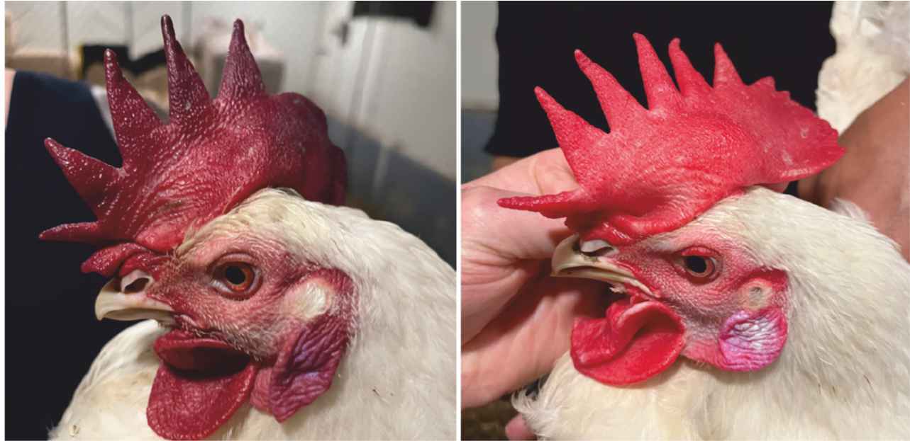
Cyanosis (purple head) is observed in breeder males, typically around 35–40 weeks of age, and is sometimes used as a management indicator (Figure 1). The condition is not limited to one breed or line and may be transient, with color changes throughout the day; cyanosis appears after feeding, then disappears during the day. Affected birds show no respiratory distress, and necropsy reveals few gross lesions except occasional flaccid hearts. Echocardiography has detected aortic insufficiency (AOI) in a significant proportion of affected males, and laboratory findings show elevated hematocrit and hemoglobin concentrations, suggesting increased blood viscosity and potential compensatory polycythemia, a condition where an overproduction of red blood cells compensates for a lack of oxygen. This information is intended to provide physiological context and support practical management decisions, rather than to imply differences between breeds or genetic lines. There is no evidence of a genetic basis for this condition; therefore, management should be the focus to reduce field incidence.
FIGURE 1: An example of affected (left) and unaffected (right) broiler breeder males.
AVIAGEN Brief: Cyanosis (Purple Head) in Broiler Breeder Males | © 2026 Aviagen
PRACTICAL MANAGEMENT OF MALE CYANOSIS
FEEDING AND PHYSICAL STRESS
Cyanosis typically occurs during periods of peak physical stress, such as feeding or mating, when oxygen demand spikes and the heart may struggle to keep pace. For this reason, feed distribution should be completed within three minutes to prevent uneven gorging and panic. Male exclusion grills must be intact to prevent males from stealing hen feed, which increases their body mass and oxygen demand while physically restricting their air sacs. In addition, verify water quality, drinker space, and water line flow rates to ensure they meet the requirements for broiler breeder males. Field observations have also suggested that behaviors such as litter consumption may contribute to transient episodes in some flocks, although the underlying mechanism remains unclear.
BODY WEIGHT AND CONDITION CONTROL
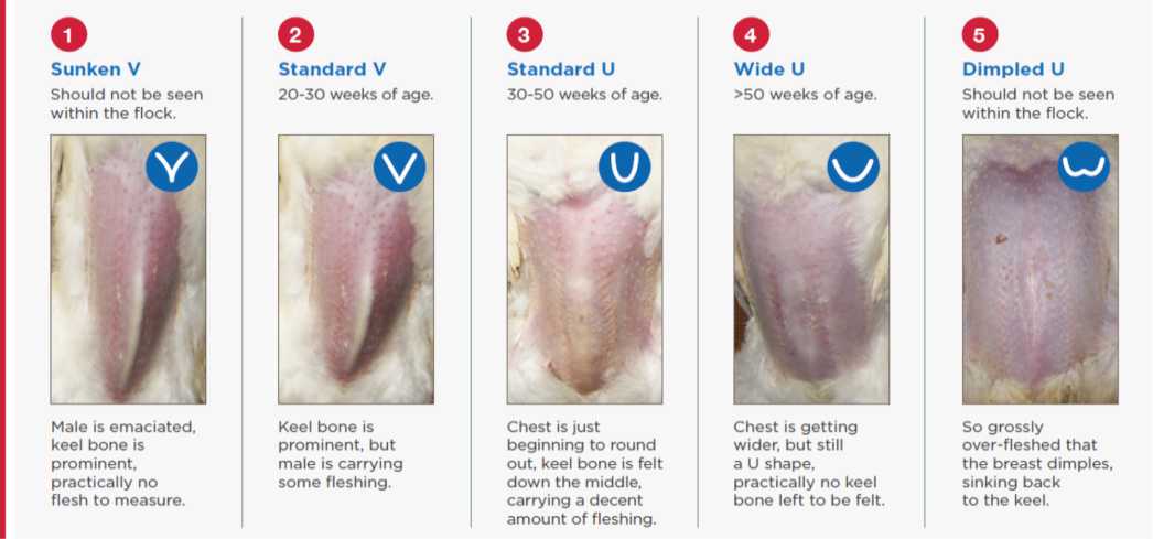
Rear males to a body weight profile that meets or slightly exceeds current breed standards, as these higher profiles help prevent conditions such as Valgus-Varus Deformity (VVD), also known as ‘X-legs’. Achieving and maintaining good body weight and uniformity in male populations is as critical as it is in females, because cyanosis occurs less frequently in well-managed, well-fed males. Over-fleshed males (with a Dimpled U-shaped breast; score 5) are at higher risk since muscle tissue is oxygen-expensive. Therefore, target a fleshing score of 2.5–3 (a Standard V or Standard U-shaped breast; score 2-3) to minimize risk (Figure 2).
FIGURE 2: Scoring system to assess bird body condition (fleshing).
VENTILATION
Oxygen demand peaks during feeding, regardless of temperature. Increase ventilation 10 minutes before and during feeding to maximize oxygen availability. Maintaining good air quality and effective air exchange is important, as suboptimal ventilation, elevated ammonia or carbon dioxide levels, and reduced oxygen availability may add to physiological stress and contribute to symptom expression, even in open-sided housing systems.
TRIAGE AND CULLING
If cyanosis is observed, monitor the bird’s recovery post-feeding. Persistent cyanosis indicates heart failure and warrants culling.
MORPHOMETRIC DOCUMENTATION OF CARDIAC ABNORMALITIES
STUDY OVERVIEW
Research by Floyd D. Wilson et al (2016) documented a high prevalence of left ventricular dilated cardiomyopathy in mature broiler breeder males, both cyanotic and clinically normal. To better contextualize these findings within commercial production, the study also incorporated comparisons to market-age broilers.
KEY FINDINGS
Both cyanotic and normal broiler breeder males showed extensive left ventricular chamber dilation, with no significant difference between groups. Older males (average 42 weeks of age) had significantly higher left and right ventricular weight-to-total heart weight ratios compared with market-age broilers (average 7 weeks of age).
Morphometric data indicated that the left ventricle chamber area-to-total ventricle area ratio increased from 3.2% in broilers to 10% in broiler breeder males; 33% of males had left ventricular volume ratios above 10%, with 13% above 20%. The right ventricle-to-total ventricle weight ratio (RV/TV) increased from 0.18 in broilers to 0.25 in males, and 36% of males exceeded the threshold for right ventricular hypertrophy, although ascites was not observed. Left ventricle wall area-to-body weight ratios were similar between age groups.
Histopathology revealed only minimal endocardiosis, with no substantial changes suggestive of dilated cardiomyopathy.
Collectively, these findings suggest that left ventricular enlargement in broiler breeder males is primarily due to dilation rather than hypertrophy, possibly linked to systemic hypertension and aortic insufficiency, and they highlight the need for further research into the relationship between cyanosis, cardiac dilation, and breeding performance.
CONCLUSIONS AND RECOMMENDATIONS
The combined evidence from pathological and morphometric studies underscores the complexity of health challenges in broiler breeder males. Cyanosis and cardiac dilation are prevalent and multifactorial, impacting both flock management and productivity. Management strategies should prioritize rapid feed distribution, strict control of body weight and uniformity, sufficient drinker space and flow rates, and enhanced ventilation during feeding. Continued research and vigilant health monitoring are essential for optimizing breeder welfare and performance.